
Journal club 25-07-11
Arrestin-biased allosteric modulator of neurotensin receptor 1 alleviates acute and chronic pain
DongYee Kim
Journal club 25-07-11 Read More »
Journal club 25-07-11 Read More »
Tsugunobu Andoh ∗, Chihiro Akasaka ∗, Kyoko Shimizu †, Jung-Bum Lee ‡, Yoko Yoshihisa †, Tadamichi Shimizu †
∗Department of Applied Pharmacology, Graduate School of Medicine and Pharmaceutical Sciences, University of Toyama, Toyama, Japan
†Department of Dermatology, Graduate School of Medicine and Pharmaceutical Sciences, University of Toyama, Toyama, Japan
‡Laboratory of Medicinal Bio-resources, Graduate School of Medicine and Pharmaceutical Sciences, University of Toyama, Toyama, Japan
https://doi.org/10.1016/j.ajpath.2019.05.017
α-Melanocyte–stimulating hormone (α-MSH) is an endogenous peptide hormone involved in cutaneous pigmentation in atopic dermatitis (AD) with severe itching. α-MSH elicits itch-related responses in mice. We, therefore, investigated whether α-MSH was involved in itching in AD. In the skin of AD patients and mice with atopy-like dermatitis, α-MSH and the prohormone convertase 2, which is the key processing enzyme for the production of α-MSH, were distributed mainly in keratinocytes. In the skin of mice with dermatitis, melanocortin receptors (MC1R and MC5R) were expressed at the mRNA level and were distributed in the dermis. In the dorsal root ganglion of mice with dermatitis, mRNAs encoding MC1R, MC3R, and MC5R were also expressed. MC1R antagonist agouti-signaling protein inhibited spontaneous scratching in mice with dermatitis. In healthy mice, intradermal α-MSH elicited itch-associated responses, which were inhibited by thromboxane (TX) A2 receptor antagonist ONO-3708. In mouse keratinocytes, α-MSH increased the production of TXA2, which was inhibited by adenylyl cyclase inhibitor SQ-22536 and Ca2+chelatorEGTA. In mouse keratinocytes treated with siRNA for MC1R and/or MC5R, α-MSH–induced TXA2 production was decreased. α-MSH increased intracellular Ca2+ ion concentration in dorsal root ganglion neurons and keratinocytes. These results suggest that α-MSH is involved in itching during AD and may elicit itching through the direct action of primary afferents and TXA2 production by keratinocytes.
Journal Club 25-07-04 Read More »
Jun Lin 12318, Qixing Nie 12418, Jie Cheng 5618, YaNi Zhong 518, Tianyao Zhang 518, Xiuying Zhang 718, Xiaoyan Ge 618, Yong Ding 12318, Canyang Niu 58, Yuhua Gao 123, Kai Wang 123, Mingxin Gao 9, Xuemei Wang 123, Weixuan Chen 10, Chuyu Yun 10, Chuan Ye 123, Jinkun Xu 123, Weike Shaoyong 123, Lijun Zhang 9, Pan Shang 56, Xi Luo 123, Zhiwei Zhang 123, Xin Zheng 9, Xueying Sha 9, Jinxin Zhang 123, Shaoping Nie 4, Xuguang Zhang 11, Fazheng Ren 12, Huiying Liu 123, Erdan Dong 81314, Xiao Yu 9, Linong Ji 7, Yanli Pang 11516, Jin-Peng Sun 56, Changtao Jiang 1231719
1Department of Immunology, School of Basic Medical Sciences, State Key Laboratory of Female Fertility Promotion, Center for Reproductive Medicine, Third Hospital, Peking University, Beijing, China2NHC Key Laboratory of Medical Immunology, Peking University, Beijing, China3Department of Physiology and Pathophysiology, Center for Obesity and Metabolic Disease Research, School of Basic Medical Sciences, State Key Laboratory of Vascular Homeostasis and Remodeling, Peking University, Beijing 100191, China4State Key Laboratory of Food Science and Resources, China-Canada Joint Lab of Food Science and Technology, Key Laboratory of Bioactive Polysaccharides of Jiangxi Province, Nanchang University, Nanchang, China5Department of Biochemistry and Molecular Biology, School of Basic Medical Sciences, Shandong University, Jinan, China6Advanced Medical Research Institute, Meili Lake Translational Research Park, Cheeloo College of Medicine, Shandong University, Jinan, China7Department of Endocrinology and Metabolism, Peking University People’s Hospital, Peking University Diabetes Centre, Beijing, China8Research Center for Cardiopulmonary Rehabilitation, University of Health and Rehabilitation Sciences Qingdao Hospital (Qingdao Municipal Hospital), School of Health and Life Sciences, University of Health and Rehabilitation Sciences, Qingdao, China9Key Laboratory Experimental Teratology of the Ministry of Education and Department of Physiology, School of Basic Medical Sciences, Shandong University, Jinan, China10Department of Obstetrics and Gynecology, Peking University Third Hospital, Beijing, China11Shanghai Institute of Nutrition and Health, The Chinese Academy of Sciences, Shanghai, China12Department of Nutrition and Health, Beijing Advanced Innovation Center for Food Nutrition and Human Health, China Agricultural University, No. 10 Tianxiu Road, Haidian District, Beijing 100193, China13The Institute of Cardiovascular Sciences, School of Basic Medical Sciences, State Key Laboratory of Vascular Homeostasis and Remodeling, NHC Key Laboratory of Cardiovascular Molecular Biology and Regulatory Peptides, Beijing Key Laboratory of Cardiovascular Receptors Research, Health Science Center, Peking University, Beijing, China14Department of Cardiology and Institute of Vascular Medicine, Peking University Third Hospital, Beijing, China15National Clinical Research Center for Obstetrics and Gynecology (Peking University Third Hospital), Beijing, China16Beijing Advanced Center of Cellular Homeostasis and Aging-Related Diseases, Institute of Advanced Clinical Medicine, Peking University, Beijing, China17Center of Basic Medical Research, Institute of Medical Innovation and Research, Peking University Third Hospital, Beijing, China
Received 25 February 2024, Revised 2 October 2024, Accepted 8 May 2025, Available online 29 May 2025.
https://doi.org/10.1016/j.cell.2025.05.010
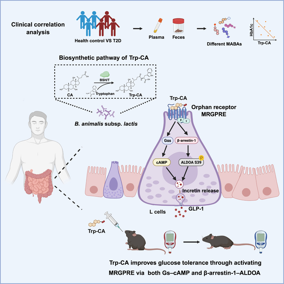
Recently, microbial amino-acid-conjugated bile acids (MABAs) have been found to be prevalent in human samples. However, their physiological significance is still unclear. Here, we identify tryptophan-conjugated cholic acid (Trp-CA) as the most significantly decreased MABA in patients with type 2 diabetes (T2D), and its abundance is negatively correlated with clinical glycemic markers. We further demonstrate that Trp-CA improves glucose tolerance in diabetic mice. Mechanistically, we find that Trp-CA is a ligand of the orphan G protein-coupled receptor (GPCR) Mas-related G protein-coupled receptor family member E (MRGPRE) and determine the binding mode between the two. Both MRGPRE-Gs-cyclic AMP (cAMP) and MRGPRE-β-arrestin-1-aldolase A (ALDOA) signaling pathways contribute to the metabolic benefits of Trp-CA. Additionally, we find that the bacterial bile salt hydrolase/transferase of Bifidobacterium is responsible for the production of Trp-CA. Together, our findings pave the way for further research on MABAs and offer additional therapeutic targets for the treatment of T2D.
Journal Club 2025.06.27 Read More »
https://doi.org/10.1016/j.phymed.2025.156564
Lina Zhou1,2+, Tunyu Jian1,+,Yan Wan2 , Rizhong Huang2 , Hailing Fang2 , Yiwei Wang2 , Chengyuan Liang1, Xiaoqin Ding1,* and Jian Chen1,2,*
1 Jiangsu Key Laboratory for the Research and Utilization of Plant Resources, Institute of Botany,
Jiangsu Province and Chinese Academy of Sciences, Nanjing 210014, China; linazhou1124@163.com (L.Z.);
jiantunyu1986@163.com (T.J.); liangcy618@cnbg.net (C.L.)
2 School of Pharmacy, Nanjing University of Chinese Medicine, Nanjing 210023, China;
wany998061@163.com (Y.W.); 20210615@njucm.edu.cn (R.H.); fanghailing2013@163.com (H.F.);
yiweiwang@njucm.edu.cn (Y.W.)
* Correspondence: dingxiaoqin@jib.ac.cn (X.D.); chenjian@jib.ac.cn (J.C.); Tel.: +86-25-8434-7104 (X.D. & J.C.);
Fax: +86-25-8434-7081 (X.D. & J.C.)
† These authors contributed equally to this work.
Journal Club 2025.06.20 Read More »
Meningeal regulatory T cells inhibit nociception
In female mice
Élora Midavaine1, Beatriz C. Moraes1, Jorge Benitez1, Sian R. Rodriguez1, Joao M. Braz1, Nathan P. Kochhar1, Walter L. Eckalbar1, Lin Tian2, Ana I. Domingos3, John E. Pintar4,
Allan I. Basbaum1*†, Sakeen W. Kashem5,6
Pain prevalence is higher in women across multiple conditions, and chronic pain severity
is frequently altered during gender affirming hormonal therapy (1). Although
there is evidence that T cells contribute to sexually dimorphic pain processing, the exact
mechanisms remain unclear (2). Regulatory T cells (Treg cells) are a subset of CD4+ T cells
defined by the expression of the master transcriptional regulator FOXP3, which is encoded
by a gene found on the X chromosome. In addition to their critical function in restraining
inflammation, Treg cells are major contributors of tissue reparative and supportive functions
(3, 4). However, it is not known whether and how Treg cells directly alter neuronal activity to
modulate nociception, independently of their immunomodulatory functions (5, 6). In this
study, we examined the role of Treg cells in regulating pain sensing in mice.
Journal Club 2025. 06. 13 Read More »
Author links open overlay panelYao Chen a†, Ziyuan Tang a†, Zhiyao Han a, Mingyang Wang a, Xinran Li a, Luying Lai a, Pingzheng Zhou a,b, Fang Wang c,d, Fengxian Li a,e,**,‡
aDepartment of Anesthesiology, Zhujiang Hospital, Southern Medical University, Guangzhou, China
bGuangdong Provincial Key Laboratory of New Drug Screening, School of Pharmaceutical Sciences, Southern Medical University, Guangzhou, China
cDepartment of Dermatology, Dermatology Hospital, Southern Medical University, Guangzhou, China
dGuangdong Provincial Key Laboratory of Brain Function and Disease, Guangzhou, China
eKey Laboratory of Mental Health of the Ministry of Education, Guangdong Province Key Laboratory of Psychiatric Disorders, Southern Medical University, Guangzhou, China
Received 7 September 2024, Revised 12 February 2025, Accepted 15 February 2025, Available online 16 February 2025, Version of Record 21 February 2025.
Atopic dermatitis (AD) is a chronic, itchy, and inflammatory skin disease. The neuroimmune concept of itch involves aberrant immune responses and neural activities. Chinese herbal medicine has been demonstrated to alleviate AD symptoms, but the underlying mechanisms remain not fully understand.
Chushizhiyang (CS) ointment is a topical treatment consisting of Chinese herbal ingredients. We aimed to study the underlying mechanism of CS on treating AD.
To investigate the therapeutic efficacy of CS, we utilized a well-established atopic dermatitis mouse model, administering CS ointment topically to the ears. To unravel the underlying mechanisms, we employed a multifaceted approach, including behavioral assay, network pharmacology analysis, RNA-sequencing analysis, neural tracing, and calcium imaging. Additionally, transient receptor potential (TRP) M8-deficient mice were employed to validate the specific targets of CS.
By employing a murine model of AD-like disease, we found that CS ointment can reduce skin inflammation and inhibit scratching behavior. Importantly, its capacity to alleviate itch-induced scratching surpasses that of topical steroid, a positive control treatment. The RNA-sequencing analysis of the affected skin revealed that the differentially expressed genes were enriched in neuroactive pathways that include ion channels particularly TRPM8. Calcium imaging demonstrated that CS ointment is capable of activating TRPM8-positive sensory neurons. Using transgenic animals, we found that CS ointment exhibited its anti-inflammatory or anti-pruritic effects only when TRPM8 is functional intact. Additionally, CS treatment reduced neuronal activities in wild-type, rather than TRPM8-compromised animals.
Our findings suggest that topical Chinese herbals participate in neuroimmune mechanisms for AD-like disease via TRPM8.
Atopic dermatitis, TRPM8, Pruritus, Inflammation, Chinese herb
AD, atopic dermatitis; TRPM8, transient receptor potential M8; CS, Chushizhiyang ointment; EtOH, ethanol; HPLC, high performance liquid chromatography; TCMSP, traditional Chinese medicine systems pharmacology database; PPI, protein-protein interaction; GO, Gene Ontology; KEGG, Kyoto Encyclopedia of Genes and Genomes; IL-4, interleukin-4; IL-13, interleukin-13, IL-31, interleukin-31; IL-33, interleukin-33; CCL17, C-C Motif Chemokine Ligand 17; TSLP, thymic stromal lymphopoietin; eGFP, enhanced greed fluorescence protein; FG, fluoro-gold; DRG, dorsal root ganglion; TG, trigeminal ganglion; pERK, phosphorylated extracellular signal-regulated kinase;
Journal Club 2025. 05. 30 Read More »
Hanbin Lee 1,2, Chigusa Nakahashi-Oda 1,3,�, Wenxin Lyu 1,2, Mamoru Tanaka1,4,
Akiyoshi Rai1,5, Yoichi Muramoto 1,5, Yaqiu Wang1,2, Seiya Mizuno6, Kazuko Shibuya 1,3, and
Akira Shibuya 1,3,7,�
Abstract
Mast cells (MCs) play a central role in allergic immune responses. MC activation is regulated by several inhibitory immunoreceptors. The CD300
family members CD300a and CD300lf recognize phospholipid ligands and inhibit the FcεRI-mediated activating signal in MCs. While CD300a
binds to phosphatidylserine (PS) to inhibit MCs activation, CD300lf function is less clear due to its ability to bind with ceramide and PS.
Moreover, it also remains blurring whether CD300a and CD300lf function independently, cooperatively, or by interfering with each other in regulating
MC activation. Using imaging and flow cytometric analyses of bone marrow-derived cultured MCs (BMMCs) from wild-type (WT),
Cd300a–/–, Cd300lf–/–, and Cd300a–/–Cd300lf–/– mice, we show that CD300lf and CD300a colocalized with PS externalized to the outer leaflet of
the plasma membrane with a polar formation upon activation, and CD300lf cooperates with CD300a to inhibit BMMCs activation. CD300lf also
colocalized with extracellular ceramide in addition to the internal PS on the cell surface, which results in stronger inhibition of MC activation than
CD300lf binding to PS alone. Similarly, although both Cd300a–/– and Cd300lf–/– mice showed decreased rectal temperatures compared with WT
mice in the model of passive systemic anaphylaxis, Cd300a–/–Cd300lf–/– mice showed lower rectal temperature than either Cd300a–/– or
Cd300lf–/– mice. Our results demonstrate the cooperativity of multiple inhibitory receptors expressed on MCs and their regulatory functions
upon binding to respective ligands.
Keywords: CD300a, CD300lf, ceramide, mast cells, phosphatidylserine
Journal Club 2025.05.23 Read More »
Mast Cells Initiate Type 2 Inflammation through Tryptase Released by MRGPRX2/MRGPRB2 Activation in Atopic Dermatitis
https://doi.org/10.1016/j.jid.2023.06.201
Tao Jia 13, Delu Che 123, Yi Zheng 1, Huan Zhang 1, Yaxiang Li 1, Tong Zhou 1, Bin Peng 1, Xueshan Du 1, Longfei Zhu 1, Jingang An 1, Songmei Geng 1
1Department of Dermatology, Northwest Hospital, The Second Hospital Affiliated to Xi’an Jiaotong University, Xi’an, China
2Center for Dermatology Disease, Precision Medical Institute, Xi’an, China
Keywords
AD: Atopic Dermatitis, ADI: Atopic Dermatitis Index, MC: Mast Cell, TSLP: Thymic Stromal Lymphopoietin
Journal Club 2025.04.25 Read More »
http://dx.doi.org/10.1097/j.pain.0000000000003540
Journal club: 2025.04.18 Read More »
Xiaohong Jin, Zimeng Chen, Dan Yu, Qianhui Jiang, Zhuobin Chen, Bin Yan, Jing Qin, Yong Liu, Junwen Wang
Bioinformatics, Volume 41, Issue 1, January 2025, btae708, https://doi.org/10.1093/bioinformatics/btae708
Published:
25 November 2024
Motivation
Peptides and their derivatives hold potential as therapeutic agents. The rising interest in developing peptide drugs is evidenced by increasing approval rates by the FDA of USA. To identify the most potential peptides, study on peptide-protein interactions (PepPIs) presents a very important approach but poses considerable technical challenges. In experimental aspects, the transient nature of PepPIs and the high flexibility of peptides contribute to elevated costs and inefficiency. Traditional docking and molecular dynamics simulation methods require substantial computational resources, and the predictive accuracy of their results remain unsatisfactory.
Results
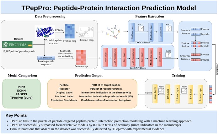
To address this gap, we proposed TPepPro, a Transformer-based model for PepPI prediction. We trained TPepPro on a dataset of 19,187 pairs of peptide-protein complexes with both sequential and structural features. TPepPro utilizes a strategy that combines local protein sequence feature extraction with global protein structure feature extraction. Moreover, TPepPro optimizes the architecture of structural featuring neural network in BN-ReLU arrangement, which notably reduced the amount of computing resources required for PepPIs prediction. According to comparison analysis, the accuracy reached 0.855 in TPepPro, achieving an 8.1% improvement compared to the second-best model TAGPPI. TPepPro achieved an AUC of 0.922, surpassing the second-best model TAGPPI with 0.844. Moreover, the newly developed TPepPro identify certain PepPIs that can be validated according to previous experimental evidence, thus indicating the efficiency of TPepPro to detect high potential PepPIs that would be helpful for amino acid drug applications.
Availability and implementation
The source code of TPepPro is available at https://github.com/wanglabhku/TPepPro.
Journal club: 2025.04.11 Read More »